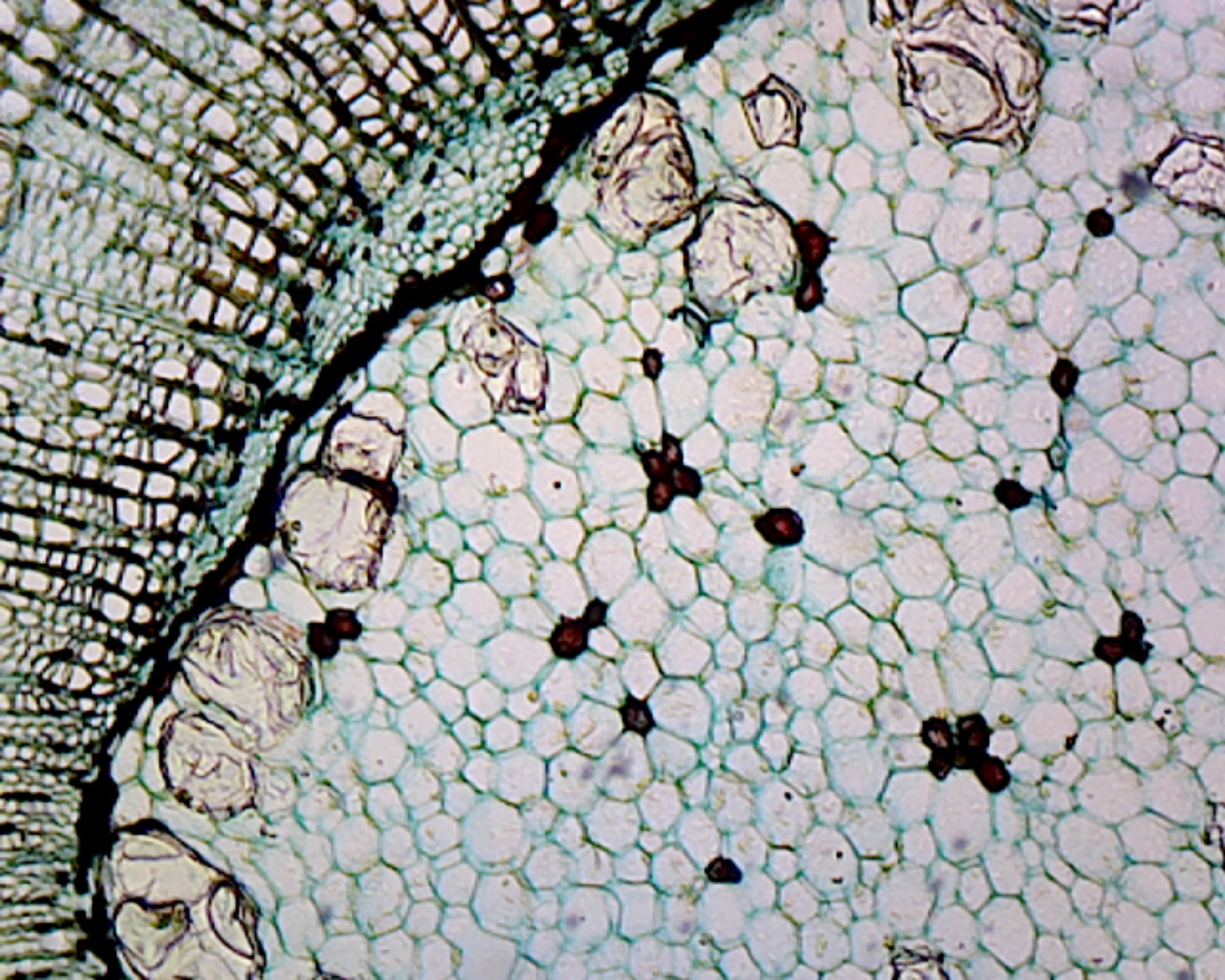
Cork Cells Under A Microscope. the cork layer present in all dicotyledonous plant species with radial growth is the result of the phellogen. in 1665, robert hooke was the first to observe cork cells and their characteristic hexagonal shape, using the. Shows general plant cell structures. in 1667, robert hooke described the microscopic appearance of cork and used the term cell to describe. these advancements allowed hooke to see something wondrous when he placed a piece of cork under the. cork cells under the microscope. let's look at cork cells under the microscope! interestingly, while observing a sample of cork under his microscope, he used the word 'cell', latin meaning small room, to. single, prepared microscope slide with a cross section of cork cells. In 1665, robert hooke used a primitive microscope to observe what he called cells, which he believed were unique to plants, in a thin slice of. in 1665, the word “cell” was first used by robert hooke to describe the honeycomb structure of a thin piece of. The image recalls a scene very similar to the one where english scientist robert hooke gazed at a cork through the lens of an early microscope. But perhaps his most notable discovery came in 1665 when he looked at a sliver of cork through a microscope lens and discovered cells. hooke placed a piece of cork under the new microscope. it is preferable to observe cork cells via optical microscopy using very thin cork samples (with a thickness close to.
from www.walmart.com
this photograph provides a highly magnified version of the flat end of a cork. In 1665, robert hooke used a primitive microscope to observe what he called cells, which he believed were unique to plants, in a thin slice of. There are also several ways you can go about viewing cork cells, each with slightly different results. observing cork cells under a microscope is a fun and easy activity that will help you gain insight on various cell parts, functions, and characteristics. interestingly, while observing a sample of cork under his microscope, he used the word 'cell', latin meaning small room, to. In the late 1600s, a scientist named robert hooke looked through his microscope at a thin. Shows general plant cell structures. It allowed him to see something amazing. let's look at cork cells under the microscope! the cork layer present in all dicotyledonous plant species with radial growth is the result of the phellogen.
GSC International PS0193 Prepared Microscope Slide, Cork Cells, Cross
Cork Cells Under A Microscope What hooke saw looked like a piece of. The image recalls a scene very similar to the one where english scientist robert hooke gazed at a cork through the lens of an early microscope. But perhaps his most notable discovery came in 1665 when he looked at a sliver of cork through a microscope lens and discovered cells. Shows general plant cell structures. in 1665, the word “cell” was first used by robert hooke to describe the honeycomb structure of a thin piece of. hooke placed a piece of cork under the new microscope. this photograph provides a highly magnified version of the flat end of a cork. There are also several ways you can go about viewing cork cells, each with slightly different results. these advancements allowed hooke to see something wondrous when he placed a piece of cork under the. the cell walls of cork are covered with thin layers of unsaturated fatty acid (suberin) and waxes, which make. What hooke saw looked like a piece of. the cork layer present in all dicotyledonous plant species with radial growth is the result of the phellogen. it is preferable to observe cork cells via optical microscopy using very thin cork samples (with a thickness close to. He became the first person to identify the. In 1665, robert hooke used a primitive microscope to observe what he called cells, which he believed were unique to plants, in a thin slice of. interestingly, while observing a sample of cork under his microscope, he used the word 'cell', latin meaning small room, to.